Wednesday, July 8, 2020

Mass Spectrometry, Class12

Mass spectrometry is a powerful analytical technique used to quantify known materials, to identify unknown compounds within a sample, and to elucidate the structure and chemical properties of different molecules. The complete process involves the conversion of the sample into gaseous ions, with or without fragmentation, which are then characterized by their mass to charge ratios (m/z) and relative abundances.

Basic Principle

A mass spectrometer generates multiple ions from the sample under investigation, it then separates them according to their specific mass-to-charge ratio (m/z), and then records the relative abundance of each ion type.

Components

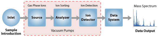
The instrument consists of three major components:
  1. Ion Source: For producing gaseous ions from the substance being studied.
MALDI -TOF:  Matrix assisted laser desorption ionization-time of flight

ESI: Electrospray Ionization
MALDI-MS and ESI-MS procedures. a In MALDI-MS, samples are ...

  1. Analyzer: For resolving the ions into their characteristics mass components according to their mass-to-charge ratio.
  2. Detector System: For detecting the ions and recording the relative abundance of each of the resolved ionic species


  1. With all the above components, a mass spectrometer should always perform the following processes:
    1. Produce ions from the sample in the ionization source.
    2. Separate these ions according to their mass-to-charge ratio in the mass analyzer.
    3. Eventually, fragment the selected ions and analyze the fragments in a second analyzer.
    4. Detect the ions emerging from the last analyzer and measure their abundance with the detector that converts the ions into electrical signals.
    5. Process the signals from the detector that are transmitted to the computer and control the instrument using feedback
the mass spectrometer - how it works


Google form, Test Ch-4, HEAT Class 7

FIBRE TO FABRIC , CH 3, CLASS 7 (Revision Notes)

Sunday, July 5, 2020

2D Gel Electrophoresis,Class12 Ch 2


This technique was developed by O'Farrel in 1975

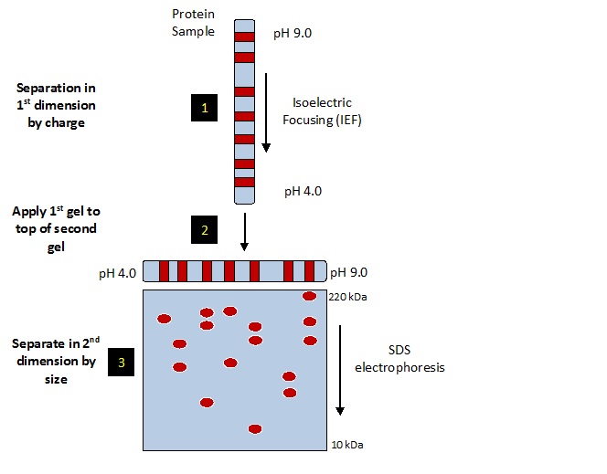
Principle: • The method resolves proteins in a protein cocktail in the form of a twodimensional protein map based on their size and charge.

Objective:
 • First dimension: Separation according to proteins isoelectric points (pI)
 • Second dimension: Separation according to molecular weight by SDS PAGE

Procedure: •Proteins are separated by their intrinsic charges in a solution which solublizes, denatures and dissociates all the polypeptide charges.

Ist dimension : Isoelectric focusing is used to separate polypeptide chains on the principal of pI.
Isoelectric point (pI) of a protein is the pH at which the protein has a net charge equal to zero. A protein does not move in an electric field when it is at its isoelectric point.

IInd dimension: The polypeptides are separated electrophoretically in polyacrylamide gel which as a pH gradient.

First dimension of the 2D gel electrophoresis is established by the movement of each protein to a position, which corresponds to its isoelectric point in a narrow tube. • This narrow tube is again subjected to electrophoresis in a direction, which is at right angle to the direction that has been used for isolectric focusing. In the second electrophoresis SDS is used to separate the proteins according to their sizes. The second dimension is established by the migration of the separated protein to its discrete spot on the gel.

After fractionation a specific protein can be identified on the gel by radioactive isotope, a detectable enzyme or fluorescent dye.
Method for HCP Analysis using 2D Gel Electrophoresis
ISO ELECTRIC FOCUSSING-
Separation of the proteins by isoelectric point is called isoelectric focusing (IEF). When a gradient of pH is applied to a gel and an electric potential is applied across the gel, making one end more positive than the other. • At all pH values other than their isoelectric point, proteins will be charged. If they are positively charged, they will be pulled towards the negative end of the gel and if they are negatively charged they will be pulled to the positive end of the gel. The proteins applied in the first dimension will move along the gel and will accumulate at their isoelectric point; that is, the point at which the overall charge on the protein is 0 (a neutral charge). 

SDS - PAGE
 In separating the proteins by mass, the gel treated with sodium dodecyl sulfate (SDS) along with other reagents (SDS-PAGE in 1-D). This denatures the proteins (that is, it unfolds them into long, straight molecules) and binds a number of SDS molecules roughly proportional to the protein's length. Because a protein's length (when unfolded) is roughly proportional to its mass, Since the SDS molecules are negatively charged, the result of this is that all of the proteins will have approximately the same mass-to-charge ratio as each other.The gel therefore acts like a molecular sieve when the current is applied, separating the proteins on the basis of their molecular weight with larger proteins being retained higher in the gel and smaller proteins being able to pass through the sieve and reach lower regions of the gel.


https://www.youtube.com/watch?v=JqFmnxGMc2g


Friday, July 3, 2020

Chymotrypsin, a proteolytic enzyme (Questions)

1. Why proteins are secreted as zymogens?
2. Name the inactive and active form of  chymotrypsin.
3. How many polypeptides are there in active chymotrypsin?
4. Why the activation of chymotrypsin is termed as in - situ activation?
5. Name different enzymes required for in-situ activation of chymotrypsin?
6. What do you mean by catalytic triad in relation with chymotrypsin?
7. Why does ser 195 becomes acidic?
8. Name the different enzymes that involves the acidic serine in its catalysis?
9. Explain in detail about the activity of chymotrypsin with the help of diagram.

Thursday, July 2, 2020

MCQs of RDT Ch-1, Class 12

An enzyme catalysing the removal of nucleotides from the ends of
DNA is:
a. endonuclease
b. exonuclease
c. DNA ligase
d. Hind – II
The transfer of genetic material from one bacterium to another through
the mediation of a vector like virus is termed as:
a. Transduction
b. Conjugation
c. Transformation
d. Translation
Which of the given statement is correct in the context of observing DNA
separated by agarose gel electrophoresis?
a. DNA can be seen in visible light
b. DNA can be seen without staining in visible light
c. Ethidium bromide stained DNA can be seen in visible light
d. Ethidium bromide stained DNA can be seen under exposure to
UV light
An antibiotic resistance gene in a vector usually helps in the selection of:
a. Competent cells
b. Transformed cells
c. Recombinant cells
d. None of the above
Significance of ‘heat shock’ method in bacterial transformation is to
facilitate:
a. Binding of DNA to the cell wall
b. Uptake of DNA through membrane transport proteins
c. Uptake of DNA through transient pores in the bacterial cell wall
d. Expression of antibiotic resistance gene
The role of DNA ligase in the construction of a recombinant DNA
molecule is:
a. Formation of phosphodiester bond between two DNA fragments
b. Formation of hydrogen bonds between sticky ends of DNA
fragments
c. Ligation of all purine and pyrimidine bases
d. None of the above
Which of the following bacteria is not a source of restriction
endonuclease?
a. Haemophilus influenzae
b. Escherichia coli
c. Agrobacterium tumefaciens
d. Bacillius amyloli
Which of the following steps are catalysed by Taq polymerase in a PCR
reaction?
a. Denaturation of template DNA
b. Annealing of primers to template DNA
c. Extension of primer end on the template DNA
d. All of the above
A bacterial cell was transformed with a recombinant DNA that was
generated using a human gene. However, the transformed cells did not
produce the desired protein. Reasons could be:
a. Human gene may have intron which bacteria cannot process
b. Amino acid codons for humans and bacteria are different
c. Human protein is formed but degraded by bacteria
d. All of the above
Who among the following was awarded the Nobel Prize for the
development of PCR technique?
a. Herbert Boyer
b. Hargovind Khurana
c. Kary Mullis
d. Arthur Kornberg
Which of the following statements does not hold true for restriction
enzyme?
a. It recognises a palindromic nucleotide sequence
b. It is an endonuclease
c. It is isolated from viruses
d. It produces the same kind of sticky ends in different DNA
molecules
A recombinant DNA molecule can be produced in the absence of the
following:
a. Restriction endonuclease
b. DNA ligase
c. DNA fragments
d. E.coli
In agarose gel electrophoresis, DNA molecules are separated on the basis
of their:
a. Charge only
b. Size only
c. Charge to size ratio
d. All of the above
The most important feature in a plasmid to be used as a vector is:
a. Origin of replication (ori)
b. Presence of a selectable marker
c. Presence of sites for restriction endonuclease
d. Its size
While isolating DNA from bacteria, which of the following enzymes is not
used?
a. Lysozyme
b. Ribonuclease
c. Deoxyribonuclease
d. Protease
Which of the following has popularised the PCR (polymerase chain
reactions)?
a. Easy availability of DNA template
b. Availability of synthetic primers
c. Availability of cheap deoxyribonucleotides
d. Availability of ‘Thermostable’ DNA polymerase